Wilms Tumor in Children – Just Diagnosed

Diagnosing Wilms Tumor and Other Kidney Tumors
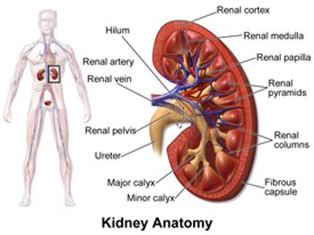
Kidney Anatomy

Wilms tumor is the most common type of kidney tumor in children, representing about 85% of diagnoses. The second most common kidney tumor is clear cell sarcoma of the kidney (CCSK). Other less common kidney tumors include renal cell carcinoma, malignant rhabdoid tumor, and congenital mesoblastic nephroma. It is important for doctors to correctly identify which tumor your child has, because each tumor is treated differently.

Typically, cases of Wilms tumor are not diagnosed until the tumor becomes quite large. Fortunately, most tumors are discovered before they metastasize (spread to other organs in the body). As with many cancers, the tumor mass can become quite large; the average weight of a newly discovered tumor is about 500 grams (one pound), which is larger than the normal kidney.

In addition to a complete medical history and physical examination, diagnostic tests are performed to evaluate the kidney tumor and look for signs of spread to other areas of the body.

  • Laboratory Tests: Certain blood tests are done to count the number of white blood cells, to look for anemia (low number of red blood cells) and to measure certain chemicals and salts in the blood that give clues about how well the kidneys are working
  • Urinalysis: A urine sample is examined using various testing methods for the presence of blood and other substances
  • Imaging studies: Many technologies are available to get a clear picture of the tumor and surrounding structures, these include:

These tests also enable the treating doctors and surgeons to select the best approach for treatment because they show where the tumor is located. The diagnosis is usually confirmed by the surgical removal of all or part of the tumor when a pathologist examines it under a microscope. In some cases, treatment with chemotherapy will be started to shrink the tumor initially and make removal safer and more effective at a later time.

Prognosis and Survival

The prognosis (chance of recovery) for children with Wilms tumor is generally very good. Currently, the great majority of children with Wilms tumor are cured. The overall five-year survival rate for children with Wilms tumor is approximately 90%. That means that 9 out of 10 children with Wilms tumor will live at least five years after their cancer is diagnosed. The five-year survival mark is the point at which a patient is considered “cured” since these tumors almost never recur after that time.

About 92% of Wilms tumors have favorable histology, meaning there is no anaplasia (distortion of the nuclei of the cancer cells), and that the chance of a cure is excellent. In very few cases, a tumor will have an unfavorable histology, meaning that the cells are abnormal and that the nuclei of the cancer cells (the part that contains the cells’ DNA) are large and distorted. This is called anaplasia. Anaplasia is more resistant to chemotherapy treatment, and therefore the chances of a cure are lower.

Wilms Tumor Staging

After making a diagnosis of Wilms tumor, doctors need to assess how advanced the tumor is, including:

  • How large it is,
  • How far it has spread, and
  • Whether it has invaded other tissues.

This is called “staging” the tumor. Staging allows the cancer care team to identify the best treatment approach using a combination of chemotherapy, surgery and radiation therapy. It will also provide information about the outlook for cure.

The staging system used to describe the extent of spread of Wilms tumors was developed by the National Wilms Tumor Study Group (NWTS). The NWTS system describes Wilms tumor stages using Roman numerals I through V (1 to 5).

Stage I

The tumor is only in the kidney, and was removed completely by surgery. The cancer has not grown into blood and lymph vessels next to the kidney. The tissue layer (the capsule) that surrounds the cancer was not broken. About 40% of all Wilms tumors are stage I.

Stage II

The cancer has grown beyond the kidney, but was completely removed surgically without any apparent cancer left behind. One or more of the following features may be present:

  • The cancer extended beyond the kidney into nearby fatty tissue, but was removed completely
  • Cancer extended into blood vessels near the kidney, but was removed

About 20% of all Wilms tumors are stage II.

Stage III

This stage refers to Wilms tumors that partially remain in the abdomen after surgery. One or more of the following features may be present:

  • Cancer has spread to lymph nodes in the abdomen or pelvis, but not to more distant lymph nodes, such as those within the chest
  • Cancer cells are present at the edge of the tissue removed by surgery, indicating that some of the cancer still remains after surgery
  • Cancer “spilled” into the abdominal cavity before or during surgery
  • The cancer invaded nearby vital structures, and the surgeon could not remove it completely
  • Deposits of tumor (tumor implants) are found along the lining of the abdominal cavity

About 20% of all Wilms tumors are stage III.

Stage IV

The cancer has spread through the bloodstream to other organs far away from the kidneys, such as the lungs, liver, bone or to lymph nodes. About 10% of all Wilms tumors are stage IV.

Stage V

Tumors are in both kidneys at the time of diagnosis. Most of the time Stage V tumors are tumors that have developed from primitive, abnormal tissue on both kidneys and do not represent spread from one kidney to the other. The treatment is based on the kidney with the more advanced stage after surgery.

About 5% of all Wilms tumors are stage V.

Pin It on Pinterest

Scroll to Top