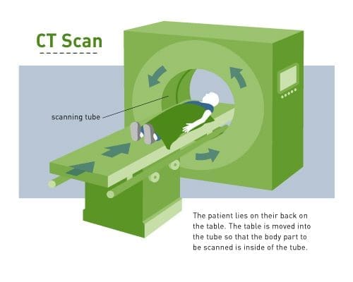
A computerized tomography (CT) scan combines a series of X-ray images taken from different angles around your body and uses computer processing to create cross-sectional images (slices) of the bones, blood vessels, and soft tissues inside your body.
The above video demonstrates a CT scan for a child undergoing cancer treatment. This video was designed to educate parents and caregivers by showing actual patients receiving treatment. Our intent with this video is to help you – and your child if you chose to show it to them – understand what will happen during this procedure.
Performing the Procedure
 CT scans are not invasive and are performed in the radiology department of a hospital. CT scans allow doctors to look not only at a person’s bones but also at soft tissue and blood vessels. CT scans are often used for diagnosing cancer because doctors can see the exact size and location of a tumor inside the patient’s body.
Sometimes, CT scans are performed using contrast dye, to better examine a specific part of the body by making it appear opaque. When this is needed, the patient is given a thick, white liquid to drink before the test. Once enough time has passed for the contrast to reach the intestines, a CT scan can be performed. A CT scan does not hurt, but it may be uncomfortable or difficult for children to very lie still for a while. Some CT scans take only a few moments, while others can take more than an hour. The length of time depends on what part of the body is being imaged. For very young children, mild sedation is often used to help them lie still.
