Nuclear medicine scans use small amounts of radioactive dye (tracer) to highlight areas of concern, such as cancer cells or infection. Pictures can than be taken of these areas. There are many types of nuclear medicine scans, including:

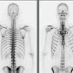
A special dye called a radioactive isotope, or tracer, is given through an IV. This tracer contains a small amount of radiation, about the same amount as an x-ray. The dye travels through the body and gathers in the area of the body being examined. When the dye has gathered in a tumor or organ, it gives off energy in the form of gamma rays. A special scanner or camera then captures images based on the gamma rays. Nuclear medicine scan pictures can detail both the function and structure of tissues and organs in the body.
The small amount of radioactive material will decay over time. It may also pass out of your child’s body through urine or stool during the first few hours or days following the test. You may be instructed to take special precautions after your child urinates, such as flushing the toilet twice and washing their hands thoroughly. Drinking plenty of water may help flush the radioactive material out of the body. Follow the instructions given to you by the nuclear medicine staff.
Nuclear medicine scans do not hurt. Allergic reactions to the tracers can occur, but they are extremely rare and are usually mild.